How Ultrasound Enables Minimally & Non-Invasive Surgery
The use of ultrasound has long been standard practice in hospitals and treatment centers throughout the world. Many people will recognize ultrasound for its imaging capabilities, which typically involves an ultrasonic scanner employing a piezoelectric transducer to generate and emit ultrasonic energy. Ultrasound can penetrate body tissue without harming it, but once it encounters a medium with a different acoustic impedance than the one it is passing through, a portion of it will ricochet off the medium boundary. This ultrasonic echo is then registered by the piezoelectric transducer and converted into an electric signal that can be used to generate an image of the matter exposed to the ultrasound.
Ultrasound in Therapeutic Medicine
Yet, the medical utilization of ultrasound extends well beyond diagnostic imaging, having expanded to include therapeutic use as well. The physical properties of ultrasound can make it an effective tool in active treatments, even enabling minimally invasive and entirely non-invasive surgical procedures for treating conditions that previously required open surgery to cure. Avoiding invasive procedures where possible is an attractive option for patients and medical practitioners alike, as it significantly reduces the risk of complications - immediate and long term - that follow from open surgery. In addition, minimally invasive and non-invasive surgeries are typically less time consuming, both in terms of time spent in the operating room and recovery time, while also being a less costly alternative without compromising the effectiveness of the treatment. Patients will also experience less scarring in the aftermath of such surgeries as open incisions can be kept to a minimum or even omitted completely.
The term ‘ultrasound’ refers to sound waves that oscillate at a frequency of 20,000 Hertz (Hz) or above which is beyond the upper limit of human hearing - with the unit ‘Hertz’ describing the number of full oscillation cycles per second. As a sound’s frequency increases, its wavelength will simultaneously shorten, and with a shorter wavelength, it becomes possible to precisely focus the sound at small target locations, enabling a high degree of energy density. With a high-power piezoelectric transducer, high-intensity focused ultrasound (HIFU) can be delivered, where the sheer intensity of the ultrasonic energy will serve to stimulate, disrupt or even destroy the targeted cells in the body. Depending on the procedure, this can be done with few to no surgical incisions being made, thereby enabling minimally invasive and non-invasive procedures.
Histotripsy - Non-Invasive Liver Tumor Radiation
Histotripsy describes the process of crushing (from the Greek word “tripsis”) organic tissue (from the Greek “histos”), in the present sense using high-intensity focused ultrasonic waves. Particularly, the technique is applied to small-size tumors in the liver. The treatment has been approved in the US and the UK and is already showing great promise as a non-invasive alternative to open surgery.
Histotripsy targets early-stage liver tumors with focused ultrasonic energy generated by an extracorporeal piezoelectric transducer to essentially pulverize the affected tissue through a process known as ‘inertial cavitation’. The process takes advantage of the immanent gas nuclei in the cells, causing them to expand and burst. Ultrasonic waves consist of alternating compression and rarefaction cycles, and during compression, the mechanical pressure on the tumor tissue is greatly increased, squeezing it together. During the rarefaction cycle, the tissue expands, and the gas nuclei bubbles within it amplifies. Once the compression cycle hits again, the bubbles will collapse under the pressure, producing shock waves, high shear forces and sudden spikes in temperature that will disintegrate the tumor from within.
Extracorporeal Shock Wave Lithotripsy (ESWL) - Non-Invasive Kidney Stone Treatment
Lithotripsy (from the Greek “lithos”, meaning “stone”) is the process of breaking kidney stones into smaller fragments using ultrasonic shock waves so that they may pass through the urinary system. Introduced in the 1980s, lithotripsy is a common non-invasive treatment method and can often be carried out in a matter of minutes with the patient being fully awake.
Lithotripsy also involves the targeting of high-intensity focused ultrasound, but while the procedure does make use of inertial cavitation, as histotripsy does, the primary ablation mechanism is high-pressure ultrasonic shock waves. Initially, the kidney stone is located using either x-rays or ultrasound scanning. Once the stone has been pinpointed, a high-power ultrasound transducer emits a shock wave pulse which will fracture the stone upon impact. Cavitation is induced, but its effect remains secondary to the initial impact of the shock wave. The fragmented kidney stone can then pass through the urinary system, having been broken into smaller pieces. Occasionally, ultrasonic waves are subsequently used to reposition residual stone fragments, making passaging easier.
Piezoelectric Solutions for Histotripsy and Lithotripsy

 Histotripsy and lithotripsy will typically employ low-frequency ultrasound for deeper penetration. As the treatments require piezoelectric transducers capable of operating at high power levels for maximal acoustic energy output, a hard-doped PZT ceramic will be the ideal material. Hard-doped PZT exhibits low dielectric and mechanical losses as well as high mechanical quality factors, meaning that more electric energy goes into acoustic output rather than heat. A common transducer design for histotripsy and lithotripsy will consist of multiple piezoelectric plates or composites arranged in so-called ‘phased arrays’ which will allow the operator to steer and alter the focus point without having to move the device itself. Such a design can also enable the generation of multiple focus points at the same time.
| Solutions for Histotripsy and Lithotripsy | |
|---|---|
| Component | Plate, 1-3 Composite |
| Recommended Material | Pz26, Pz36 |
| Frequency Range | >400 kHz |
| Max. Composite Size | 100 x 100 mm |
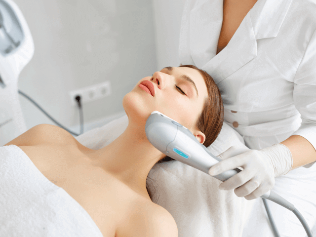
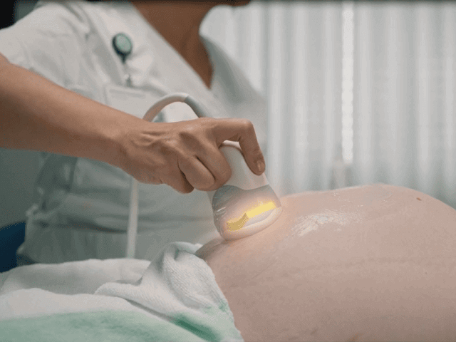
Cosmetic Dermatological Procedures - Non-Invasive Alternative to Plastic Surgery
High-intensity focused ultrasound is utilized in non-invasive cosmetic treatments that aim to reduce wrinkles and tighten the patient’s skin. Using a HIFU transducer, the dermis, the middle layer of the skin, is targeted with ultrasonic energy to stimulate its natural production of collagen and elastin. These proteins are produced by fibroblast skin cells located in the dermis and are responsible for the structural support, strength and elasticity of the skin. Over time, the density of collagen and elastin decreases, resulting in wrinkles and sagging skin. Exposing the fibroblast cells to focused ultrasound has been shown to temporarily increase their production of collagen and elastin proteins, thereby boosting the skin’s firmness and elasticity. This treatment can either´be used as a non-invasive alternative to plastic surgery, or it can be applied following an invasive cosmetic procedure, such as liposuction, to tighten loose skin.
 Piezoelectric Solutions for Cosmetic Treatments
Focused ultrasound for cosmetic treatments typically operates on the higher end of the frequency spectrum, as the sound waves only have to travel a short distance through the shallow skin tissue before reaching the target location. Employing a piezoelectric focusing bowl in a hard-doped PZT ceramic material will enable the precise concentration of ultrasonic energy, penetrating the outer skin tissue without harming it as the energy will only reach its full intensity at the transducer’s focal point.
| Solutions for Cosmetic Treatments | |
|---|---|
| Component | Focusing Bowl |
| Recommended Material | Pz26 |
| Frequency Range | 1-20 MHz |
| Max. Diameter | 100 mm |
| Penetration Depth | 0-50 mm |
Phacoemulsification - Minimally Invasive Cataract Surgery
A cataract is a cloudy area that forms in the lens of the eye as a result of the lens’ crystallin proteins breaking down. Cataracts are typically related to age, but they can also form as a result of various trauma experienced by the eye. Regardless of cause, cataracts can lead to blurry and impaired vision, increased light sensitivity and other symptoms related to sight. It is a very common condition that many people will encounter during their lifetime, but fortunately, well-established treatments exist.
Phacoemulsification is the process of breaking up cataracts by targeting them with ultrasonic energy. The procedure involves a small, self-healing incision at the edge of the cornea, through which the tip of an ultrasonic transducer is inserted. The transducer will generate ultrasonic vibrations that induces cavitation in the cataract, causing it to fall apart (emulsify). As the cataract comes apart, the fragments are simultaneous suctioned out through the same probe, and once it has been removed completely, an artificial intraocular lens (IOL) is inserted to restore the eye’s ability to focus light. Due to the minimally invasive nature of phacoemulsification, the procedure can be carried out in a matter of minutes with the patient being awake. Also, recovery time is usually short and complications few.
 Piezoelectric Solutions for Phacoemulsification
A phacoemulsification probe will typically be designed around a ring-shaped piezoelectric component. Several rings are stacked and preloaded into a handpiece, allowing high amplitude vibration in thickness mode in ideal conditions. A hard-doped PZT material with a very high mechanical quality factor is preferable due to low electrical and mechanical losses and high power generation capabilities.
| Solutions for Phacoemulsification | |
|---|---|
| Component | Ring |
| Recommended Material | NCE81 |
| Typ. Operating Frequency of Assembled Handpiece | 35-55 kHz |
Renal Denervation - Minimally-Invasive Ultrasound Treatment for Hypertension
Hypertension refers to high blood pressure, a very common condition, experienced by millions of people around the world. Blood pressure is determined by the amount of blood that the heart pumps into the arteries, and how easily the blood can pass through the arteries. The more blood the heart pumps and the narrower the arteries are, the higher the blood pressure will be. Hypertension can damage the blood vessels and the internal organs, particularly if the condition has persisted over extended periods of time. Complications include heart attacks, strokes, aneurysms, organ failure and numerous other harmful events.
In many instances, there is no single identifiable cause for high blood pressure, but risk factors include old age, obesity and lack of exercise, high-salt diet, alcohol consumption and smoking as well as genetic predisposition. Another factor that has been linked to hypertension is sympathetic overactivity, a state of excessive activity in the sympathetic nervous system. To treat sympathetic overactivity in the renal nervous system, that connects the kidneys with the central nervous system, a minimally invasive procedure utilizing ultrasound can be employed. Renal denervation, as it is called, involves inserting a catheter upon wich a small ultrasound transducer is mounted, into the main renal artery. Once in place, the transducer will deliver ultrasonic energy radially to the numerous nerves that surround the renal artery. Exposing the nerves to ultrasonic energy has been shown to reduce their excessive activity, consequently reducing blood pressure as well as alleviating hypertension.
 Piezoelectric Solutions for Renal Denervation
Renal denervation catheters will typically employ an ultrasonic transducer constructed around a very small piezoelectric tube. The geometry and polarization of this component enable 360° actuation for delivery of ultrasonic energy in all directions. A hard-doped PZT material with strong acoustic power output capabilities and a low dielectric dissipation factor is recommended for renal denervation transducers.
| Solutions for Renal Denervation | |
|---|---|
| Component | Tube |
| Recommended Material | Pz26 |
| Frequency Range | <10 MHz |
| Diameter | 1-4 mm |
Intravascular Ultrasound (IVUS) - Minimally Invasive Angioplasty Monitoring
Angioplasty is an intravascular procedure in which a catheter mounted with a deflated balloon is inserted into a partially obstructed or blocked artery in order to widen it. Once the catheter has been positioned correctly within the artery, the balloon is inflated, expanding the artery and restoring blood flow to normal. Angioplasty becomes necessary when the buildup of fats and cholesterol (known as atherosclerosis) becomes so dense that it is threatening to shut off the passage of blood that, if untreated, can lead to blood clots and heart attacks.
While not itself treating the condition, ultrasound is nonetheless an integral part of angioplasty surgery. During the procedure, an ultrasonic probe will be inserted through the balloon catheter, emitting high-frequency ultrasonic energy. As the ultrasonic waves are reflected by the artery walls and registered by the probe, a cross-sectional image of the arteries can be generated. This will enable the surgeons to find the exact location and determine the severity of the atherosclerosis within the artery and allow them to place the balloon catheter at the optimal position for the best results.
 Piezoelectric Solutions for Intravascular Ultrasound
Intravascular ultrasound will usually be in the very high end of the frequency spectrum as high spacial resolution is needed to image the artery walls. Using ultrasound in the 20-60 MHz range will provide high-resolution imaging, detailing the fine inner structures of the arteries which is needed for angioplasty procedures. A very sensitive piezoelectric material with high charge coefficients is recommended for this application as the image sharpness improves with higher sensitivity. IVUS catheters are typically designed to rotate around their own axes, meaning that a transducer built with a single piezoelectric element often will suffice for generating a 360° view of the artery. A high-frequency composite made of a PMN-PT single crystal material will provide the necessary high sensitivity and bandwidth for mapping the internal structure of the artery walls in fine detail.
| Solutions for IVUS | |
|---|---|
| Component | 1-3 High-Frequency Single Crystal Composite |
| Recommended Material |
PMN-PT Single Crystal |
| Frequency Range | 50-60 MHz |